Anatomy Of The Knee: Understanding Joint Structure
Table of Contents
- A Closer Look at the Anatomy of the Knee
- Structure and Function of the Knee Components
- Who Most Commonly Suffers From Knee Pain
- Early Warning Signs of Knee Pain
- Practical Advice for Dealing with Knee Pain
- Follow the PRICE Protocol for Acute Pain
- Apply a Hot Pack If The Condition is Chronic
- Consult An Expert for Early Diagnosis and Treatment
- Conclusion
- Frequently Asked Questions
The anatomy of the knee joint is designed in a way that provides stability for supporting body weight along with allowing movements. Knee anatomy is not just the union of two bones, rather it consists of many structures that connect the bones, separate the articular surfaces, provide cushioning, and hold the joint together. To understand the prevention of knee injuries and the healing mechanism involved you must know about the different components of the knee joint, their structure, function, and how prone each of these to getting injured. So, we’ll highlight all the aspects of the knee joint anatomy to answer the many questions related to knee pain.
A Closer Look at the Anatomy of the Knee
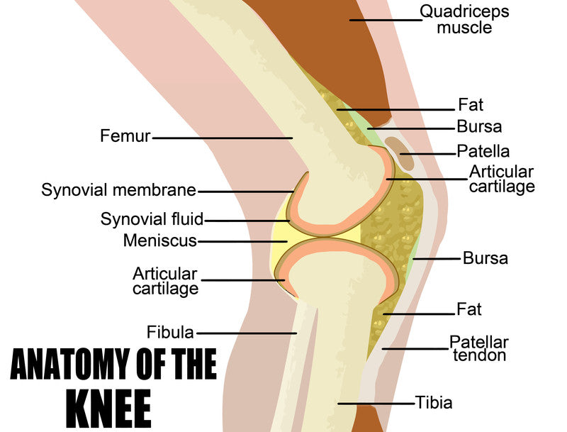
The knee joint is formed by the thigh bone (femur) and the shinbone (tibia) which has a kneecap (patella) placed above the joint that helps in protecting and moving the joint. The bones are connected by various ligaments anteriorly, posteriorly, laterally, and medially. Two menisci, the lateral and medial meniscus are present between the femur and the tibia bones to act as a cushion for reducing friction and weight bearing. All these bones, ligaments, and menisci are enclosed in a tough joint capsule completing the anatomy of the knee joint. Muscle attachments that move the joint and bursas that reduce the friction between the packed structures of the knee joint are also present either outside or inside the joint capsule.
An understanding of each of these components helps you to correlate the location of knee pain with the type of injury and the structures involved. Once you know the arrangement of structures and referred pain patterns, you can easily identify, treat, or prevent knee injuries.
For instance, pain in the front of the knee signifies a kneecap problem whereas at the back of the knee mostly indicates a baker’s cyst. Similarly, pain on the inside of the knee is an indicator of meniscus injury, but on the outside of the knee marks more towards an arthritic condition of ligament injury. Besides pain, other indicators of a knee injury include instability, giving way, and popping or cracking sounds which are more coherent from a functional point of view as a change in activity, weight gain, or sudden fall or blow might be the reason.
A closer look into the structural and functional arrangement of these components can give you an idea of the potential injuries causing knee pain, and the activities that can alter the biomechanics of the joint, as well as the best treatment options for each condition.
Structure and Function of the Knee Components
As you already know by now that the anatomy of the human knee is a complex of bones, ligaments, meniscus, muscles, tendons, and bursa, here is a detailed anatomical structure of each component along with their biomechanical functions.
Bones
The anatomy of the knee joint consists of these bones,
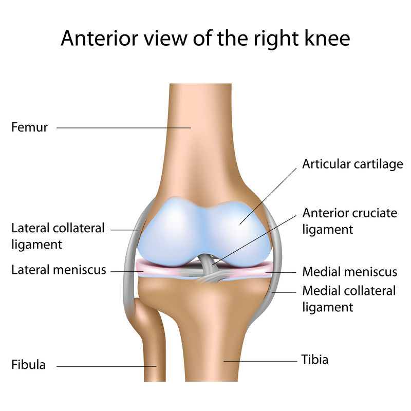
Femur: It is the thigh bone that forms the upper articular surface of the knee joint. The femoral condyles are wide and round like balls that form the joint.
Tibia: Tibia is the shin bone that forms the lower articular surface of the knee joint. The tibial condyles are also wide but are shaped like cups that serve as a platform for the rounded femoral condyles.
Patella: Patella is the kneecap that is placed right above the knee joint. It floats over the space between the tibia and femur but also forms a joint with the femur called the patellofemoral joint.
The tibia and femur form a hinge joint that moves in one plane of flexion and extension just like the hinges of a door.
Ligaments
Anterior Cruciate Ligament (ACL): It starts at the back of the lateral femoral condyle and runs downwards, inwards, and forwards to the front of the medial tibial condyle.
The function of ACL is to limit knee straightening and its injury can lead to excessive knee extension.
Posterior Cruciate Ligament (PCL): It starts at the front of the medial femoral condyle and runs downwards, outwards, and backward to the back of the tibial plateau.
It works to limit knee bending and an injury to the PCL can cause posterior displacement of the tibia while knee flexion.
Collateral Ligaments: The two collateral ligaments are the medial and lateral collateral ligaments (MCL and LCL) both located on either side of the knee joint connecting the knee bones. It protects the knee against a hit on the inside or outside of the knee. A blow on the inside of the knee can injure the LCL whereas, a blow from the outside can rupture the MCL causing pain on the respective side of the knee joint.
Menisci
The two menisci are the medial and lateral meniscus. Meniscus is formed of a tough cartilaginous structure shaped like a crescent. Each meniscus is placed on the cup-shaped tibial condyles on which the femur rests so that the weight is not directly transmitted to the bone.
It works as a cushion between the tibia and the femur.
The medial meniscus bears more weight than the lateral meniscus, and that’s why it is more prone to injury that involves twisting the leg that causes a popping and locking sensation of the knee.
Muscles and Tendons
The thigh and leg muscles are attached to or near the knee joint components. An important attachment is the Quadricep muscle of the front of the thigh. The quadriceps tendon is attached to the kneecap and runs over it to form a thick band called the patellar tendon that attaches the kneecap to the tibia.
This tendon causes many problems when installed or ruptured because it moves the patella when the knee bends or straightens.
Fat Pads and Bursa
The anatomy of the knee joint is not complete without talking about the fat pads and bursa present in the joint.
Infrapatellar Fat Pad: It is located behind the patella and acts as a cushion between the patella and the tibia and femur.
Bursa: These are fluid-filled sacs present between the bones and the tendons to cushion or reduce the friction between the structure with movement. Important bursa of the knee joint are,
- Suprapatellar bursa
- Prepatellar bursa
- Infrapatellar bursa
- Semimenbraneous bursa
An inflammation of the bursa called bursitis can occur due to overuse injuries.
Who Most Commonly Suffers From Knee Pain
Anyone can suffer from knee pain but some risk factors put a person more at risk of knee injuries. Therefore, individuals who are more prone to knee pain, especially those who commonly wear knee brace, can be identified based on the presence of these risk factors.
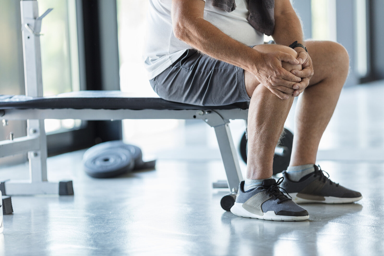
- Athletes and Active Individuals: People participating in sports have to participate in activities and movements that involve twisting and stressing the knee. Besides overuse injuries, athletes, football players, golf players, and people who do intense training can get an injury from a fall, a blow, or a twisting of the knee joint. Sprinting can cause back of knee pain.
- Older Adults: Old age is one factor for knee pain because of the degenerative changes of the cartilage that protects the joint surfaces. Knee pain due to arthritis is most common in older adults.
- Overweight and Obese Individuals: As you know the knee joint is a weight-bearing joint, so, more weight also challenges the knee even more that makes it prone to muscle, tendon, ligament, or meniscus injury.
- Occupational Factors: Jobs that involve repeated flexion and extension of the knee, or constant bending of the knee can cause pain. For instance, dancers, construction workers, drivers, or people who sit on the floor mostly have knee pain.
- Previous Knee Injuries: Because of the complexity of the knee joint, an injury of one part also weakens the other components, and often MCL tear causes medial meniscus injury followed by an ACL tear, a condition called the Unhappy Triad that can be prevented efficiently by wearing a protective knee brace.
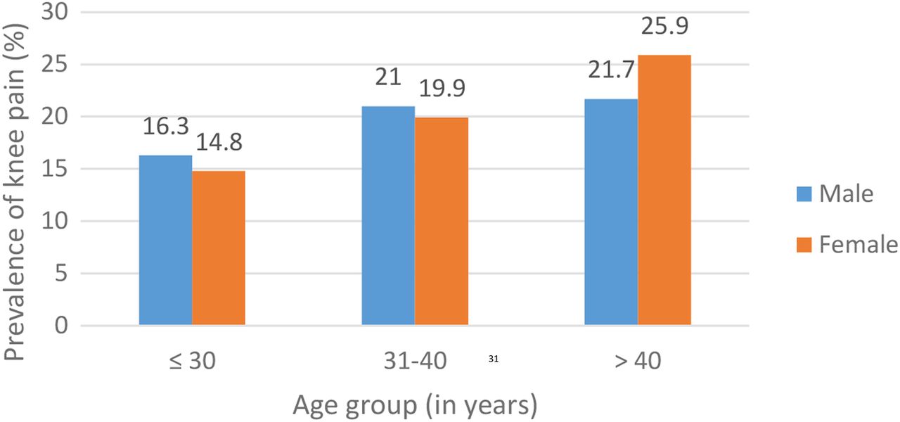
Source: https://bmjopen.bmj.com/content/6/12/e011925
Early Warning Signs of Knee Pain
Some key problems come with sudden pain while others have initial symptoms that are not painful and that’s why they are often ignored by people. These are the indicators that tell you if there is a problem with your knee joint.
- An initial discomfort that is not pain
- Popping and crunching noise with knee movement and pain on weight-bearing
- Locking of the knee
- Giving way or buckling of the knee
- Inability to straighten the knee
- Swelling, redness, and warmth
The signs and symptoms of different types of knee injuries overlap and sometimes it’s difficult to differentiate one from the other. However, with adequate attention to the symptoms and early diagnosis, you can prevent the worsening of knee pain.

Practical Advice for Dealing with Knee Pain
To get rid of knee pain, an effective way is to understand the behavior of pain and report accurate information about the location, intensity, sensation, and duration of the pain to your healthcare provider. On the basis of pain behavior and physical evaluation, your physiotherapist or other healthcare provider makes the decisions for managing your knee pain.
Here are a few tips to help you deal with knee pain,
Follow the PRICE Protocol for Acute Pain
Price stands for Protection, Rest, Icing, Compression, and Elevation which together help your knee pain to subside and let your injury heal faster.
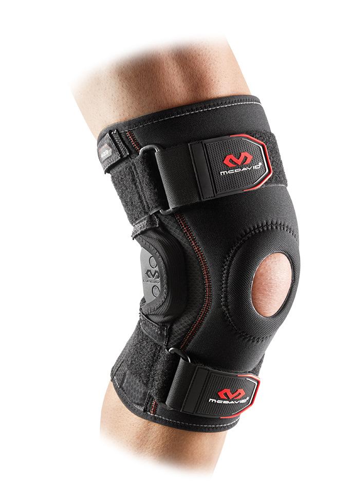
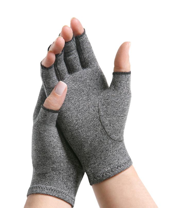
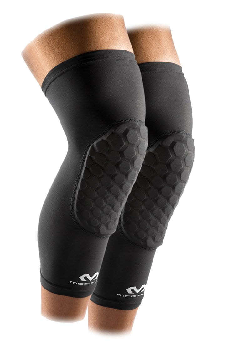
For protection and compression, you can use best knee pads instead of an immobilizer to avoid muscle wasting. Applying ice, keeping your knee elevated, and avoiding activities that stress the knee joint is crucial for recovery.
Apply a Hot Pack If The Condition is Chronic
For chronic pain and arthritic conditions, you can apply a hot pack to increase the blood flow in the region and promote healing.
Consult An Expert for Early Diagnosis and Treatment
Always consult an expert in the field for proper assessment and diagnosis. Your doctor might use physical evaluation and tests or ask for radiographic imaging to diagnose the rupture of a tendon, muscle, or meniscus tear and formulate a treatment plan.
Conclusion
The anatomy of the knee joint is complex because of the many components and their arrangement. Knowledge of the structural and functional components of knee anatomy can help you understand the causes of knee pain. You need to focus on the signs and symptoms of knee problems from the very beginning and avoid the stressors for the prevention and healing of the injury.
References:
- https://pubmed.ncbi.nlm.nih.gov/21540705/
- https://pubmed.ncbi.nlm.nih.gov/30276447/
- https://www.ncbi.nlm.nih.gov/books/NBK431067/
Frequently Asked Questions
What type of joint is the knee?
The knee is a hinge type of joint that allows movement in one dimension which is flexion and extension. It is also called a uniaxial joint because of movement only around a single axis and in a single plane.
What prevents dislocation of the knee joint anatomy?
The ligaments, muscle-tendon attachments, and the joint capsule enclose the whole anatomy of the knee joint to present it from any dislocations or instability. If these anatomical structures are weak, then a protective or supportive knee brace can aid in providing stability.
What are kneecap and knee bones called?
The kneecap is called Patella and the two other bones forming the knee joint are the shin bone called Tibia and the thigh bone called Femur.
How to relieve knee joint pain?
For relieving acute knee pain icing, rest, compression, and elevation are used with gentle stretches. For chronic knee pain, a hot pack is recommended instead of an ice pack.
What is the best painkiller for knee pain?
Taking painkillers for knee pain depends on the condition of the knee causing pain. If an inflammatory condition like tendonitis or bursitis is present then NSAIDs are used. It is always best to consult a doctor for medication instead of searching on the Internet.

Leave a comment